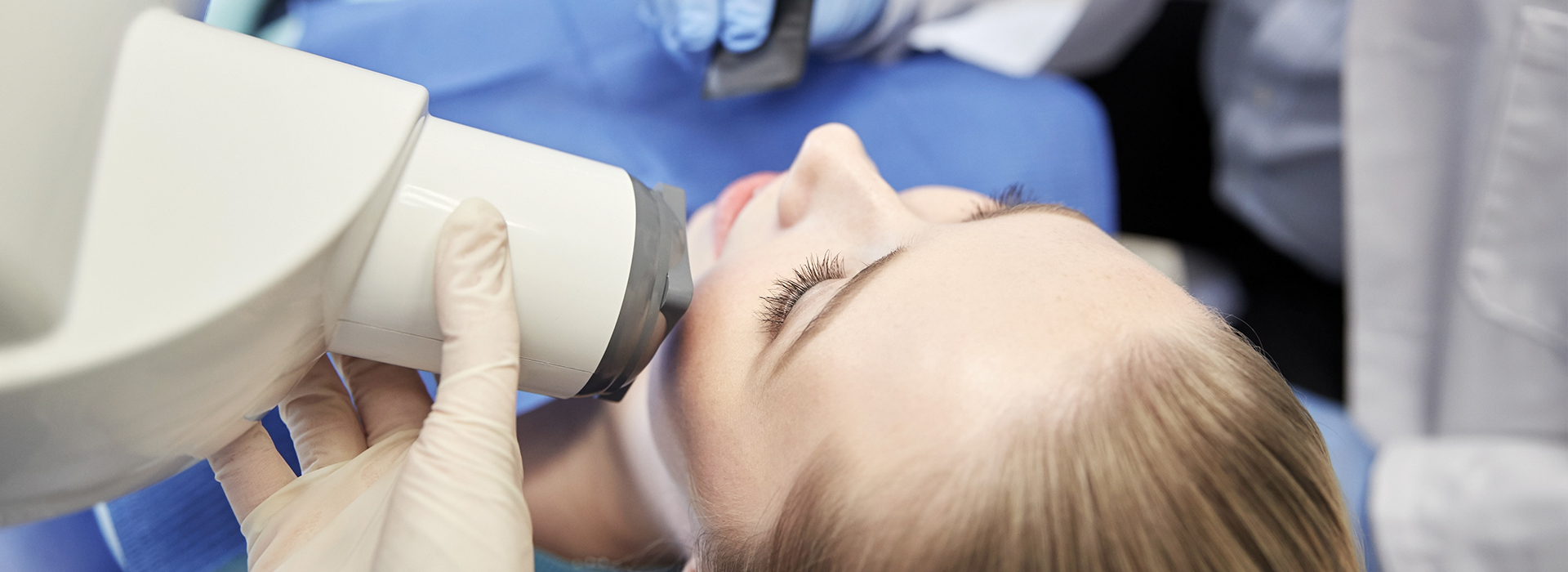
Digital radiography replaces traditional film with compact electronic sensors that capture radiographic images electronically. Instead of waiting for film to develop, the sensor immediately converts x-ray photons into a digital file that appears on a monitor within seconds. That rapid feedback helps dentists confirm image quality in real time so they can retake a shot only when necessary, reducing delays and improving diagnostic confidence during the appointment.
There are a few common sensor types used in dentistry—direct sensors that link to a computer and phosphor plate systems that are scanned into a digital file—but all share the same advantage: instant availability. Once an image is acquired, it becomes part of the patient’s digital chart, where it can be organized, compared to previous images, and enhanced for clearer interpretation. This seamless capture and storage model is a cornerstone of modern clinical workflows.
Because the image is digital from the moment it is taken, clinicians can adjust contrast, brightness, and magnification without additional radiation. These on-screen adjustments let practitioners highlight subtle details—such as early decay or minor bone changes—without subjecting patients to more exposures. The result is a smoother exam that produces usable diagnostic information quickly and reliably.
One of the most important benefits of digital radiography is its ability to reduce patient radiation exposure compared with older film-based methods. Digital detectors are more sensitive to x-rays, which means clear diagnostic images can be produced using lower doses. In everyday practice, that translates to less cumulative exposure for patients while maintaining high-quality imaging for clinical decisions.
Beyond the sensor’s sensitivity, the instant viewing capability further enhances safety. If an image is imperfect, the clinician can immediately retake it rather than waiting to discover a problem after film development. Fewer retakes mean fewer x-ray exposures overall. Additionally, modern offices follow established safety standards and imaging guidelines to ensure that every image taken is clinically justified.
For patients with concerns about radiation—such as children or people who require frequent imaging—digital radiography helps clinicians balance the need to diagnose with the imperative to minimize exposure. These considerations are part of routine care and inform how imaging is scheduled and performed for each individual patient.
Digital images often offer improved clarity and contrast compared with film, which can be especially helpful for identifying early dental conditions. On-screen tools allow dentists to zoom in on a tooth, adjust settings to reveal subtle density differences, and measure spaces with digital calipers. Those capabilities aid in detecting cavities, evaluating bone levels around teeth, and planning restorative treatments with greater confidence.
Because images are stored electronically, clinicians can compare current radiographs with prior studies side by side. This time-based view is invaluable for monitoring disease progression, assessing healing after treatment, or confirming the stability of restorations and implants. The ability to view sequential images quickly supports evidence-based treatment planning and clearer communication among the care team.
Digital files also integrate neatly with other digital technologies in the clinic, such as intraoral cameras and charting systems. When these tools work together, clinicians gain a more comprehensive picture of oral health—combining photographic detail, radiographic insight, and clinical notes—to deliver more precise and predictable care.
Digital radiography streamlines the clinical workflow from image capture to documentation. Images are automatically linked to the patient’s electronic record, eliminating manual steps like film processing and filing. That efficiency shortens appointment times, reduces administrative work, and helps the team focus on direct patient care rather than handling physical x-ray film.
Instant digital files also make collaboration simpler. When a case requires a specialist’s input—such as an oral surgeon, endodontist, or orthodontist—images can be shared electronically in a matter of moments. This quick exchange supports coordinated care and can speed decision-making for complex cases, all without requiring patients to transport physical copies or return for additional appointments.
For patients, the workflow benefits are tangible: clearer explanations during the visit, fewer follow-up calls to clarify findings, and the convenience of having all records in one secure digital system. Those efficiencies contribute to a smoother, more transparent patient experience from consultation through treatment and follow-up.
Digital radiography often improves patient comfort. Sensors and plates are slimmer and faster to position than some traditional film holders, which can reduce gagging and discomfort during intraoral imaging. Because fewer retakes are usually necessary, patients spend less time in the chair and leave their appointments with essential diagnostic information already available for discussion.
From a convenience perspective, digital images are easy to retrieve and display during consultations, which helps clinicians walk patients through findings and treatment options visually. This immediate visual feedback supports informed decision-making and helps build understanding about oral conditions and proposed interventions.
There are environmental benefits as well: digital systems eliminate the need for chemical developers, fixer solutions, and paper-based storage associated with film. Reducing these materials lowers hazardous waste and the carbon footprint tied to traditional imaging workflows, aligning dental care with broader sustainability goals.
In summary, digital radiography brings faster results, clearer images, and improved safety to routine dental care. By combining sensitive electronic sensors with integrated record systems, the office of Draper Dental can deliver more efficient appointments and more precise diagnoses while minimizing radiation and environmental waste. Contact us for more information about how digital imaging fits into your dental care and what you can expect at your next visit.
Digital radiography is a dental imaging method that uses electronic sensors to capture x-ray images and convert them into digital files almost instantly. These sensors replace traditional film and produce images that appear on a monitor within seconds, allowing clinicians to confirm image quality in real time. The immediate availability of images speeds diagnosis and reduces the need for repeat exposures.
Because images are digital from the outset, clinicians can adjust contrast, brightness, and magnification on-screen without additional radiation. Digital files also become part of the patient record, where they can be organized, compared to prior studies, and reviewed during treatment planning. This workflow improvement supports faster, more informed clinical decisions and clearer patient communication.
Unlike film x-rays, which require chemical processing, digital radiography captures radiographic information electronically and produces an image immediately. Digital detectors are more sensitive to x-rays, so clear diagnostic images typically require a lower radiation dose than film-based systems. The elimination of film processing shortens appointment times and reduces handling of hazardous chemicals and paper records.
Digital images can be enhanced and magnified on-screen to reveal subtle details that might be harder to see on film. Clinicians can compare current and past images side by side to monitor changes over time, improving long-term follow-up and evidence-based care. The streamlined storage and retrieval of digital files also simplifies collaboration with specialists when additional input is needed.
Digital radiography generally exposes patients to lower levels of radiation than traditional film x-rays because modern detectors are more efficient at capturing x-ray photons. Dentists follow the ALARA principle—keeping exposure As Low As Reasonably Achievable—by only taking images that are clinically justified and by using the lowest dose settings that produce diagnostic-quality images. Immediate image review further reduces repeat exposures by allowing clinicians to retake imperfect images right away.
For patients who require frequent imaging or for children, clinicians tailor imaging protocols to balance diagnostic needs and radiation minimization. Practices also follow established safety guidelines, use protective measures when appropriate, and document imaging decisions in the patient record. If you have specific radiation concerns, mention them to your care team so they can explain how imaging will be managed for your situation.
Common sensor types include direct intraoral sensors, which connect to a computer and deliver an image instantly, and phosphor storage plates (PSP), which are scanned after exposure to produce a digital file. Direct sensors typically use CCD or CMOS technology and provide immediate feedback, while PSP plates are flexible and work similarly to film for patient comfort but require a short scanning step. Both systems provide the instant, high-resolution images characteristic of digital radiography.
In addition to intraoral sensors, clinics may use extraoral digital systems such as panoramic sensors and cone-beam computed tomography (CBCT) for three-dimensional views when indicated. Each sensor type has strengths for specific clinical tasks, and clinicians select the appropriate modality based on the diagnostic question. Integrating these sensors into a digital workflow supports versatile, case-specific imaging strategies.
Dentists use digital x-rays to detect conditions that are not visible during a clinical exam, including early tooth decay between teeth, bone loss from periodontal disease, and the presence or position of impacted teeth. On-screen tools allow clinicians to zoom, adjust contrast, and take measurements with digital calipers to evaluate anatomy and pathology more precisely. These image enhancements often reveal subtle changes earlier than film alone could, supporting earlier intervention.
Digital radiographs also facilitate monitoring over time by enabling side-by-side comparisons with prior images to assess disease progression or healing after treatment. This chronological view is valuable for tracking bone levels around teeth, the integrity of restorations, and the outcome of endodontic therapy. Clear visual evidence helps clinicians make measured treatment recommendations and explain findings to patients more effectively.
Yes. Digital radiographs provide precise information about tooth structure, root morphology, and bone levels that are essential for planning implants, crowns, and endodontic procedures. Measurements and enhanced views allow clinicians to evaluate available bone, identify root canal anatomy, and assess the fit and margins of existing restorations. When combined with other digital tools, radiographs support predictable, evidence-based planning.
For implant planning or complex restorative cases, clinicians may combine intraoral radiographs with three-dimensional imaging like CBCT to obtain a comprehensive view of anatomy and spatial relationships. Digital workflows also make it easier to share imaging and treatment plans with labs and specialists for coordinated care. The result is more accurate planning, fewer surprises during treatment, and better long-term outcomes.
Digital dental images are typically stored within an electronic health record or imaging system using standardized formats that support interoperability and secure transfer. At Draper Dental, images are linked to each patient's chart so clinicians can retrieve, compare, and annotate studies as part of routine care. Secure systems use encryption, access controls, and regular backups to protect patient information and ensure records remain available when needed.
Clinics follow legal and regulatory requirements for patient privacy and data security, including established health information privacy standards. Authorized staff access images for clinical purposes only, and systems often maintain logs to track who viewed or modified records. If patients have questions about how their images are stored or shared, the practice can explain the specific safeguards in place.
During a digital x-ray appointment, a clinician or assistant will place a small sensor or phosphor plate inside your mouth for intraoral images or position an external sensor for panoramic views, then take a brief exposure that typically lasts only a fraction of a second. The process is quick and repeatable, and many patients notice less discomfort compared with older film holders because modern sensors and plates tend to be slimmer. After capture, the image appears on a monitor for immediate review with the clinician.
Depending on the findings, the clinician may adjust image settings, compare current images with prior studies, or take additional views if a different angle is needed for diagnosis. Protective measures, such as thyroid collars or lead aprons, are used when appropriate, and staff will follow safety protocols to minimize radiation exposure. If you have concerns about comfort or safety, mention them before imaging so the team can accommodate your needs.
Digital radiographs enable clinicians to show patients clear, enlarged images on a screen and point out areas of concern in real time, which improves understanding of diagnoses and treatment options. Visual explanations help patients see the same evidence the clinician uses, making discussions about care more transparent and easier to follow. Side-by-side comparisons with prior images also illustrate change over time, reinforcing the rationale for recommended care.
For coordination with specialists, digital files can be shared electronically, speeding referrals and collaborative treatment planning without requiring physical films. Faster exchanges reduce delays in starting specialty care and allow the referring dentist to participate in decision-making alongside consultants. These communication efficiencies support more coordinated, timely, and patient-centered care.
Draper Dental uses digital radiography to deliver faster, more precise diagnostic information while reducing radiation exposure and environmental waste associated with film processing. Digital systems integrate with the practice's patient records to streamline workflows, enable side-by-side comparisons of studies, and support clearer explanations during consultations. These capabilities help clinicians diagnose sooner and plan treatment with greater confidence.
By adopting modern imaging technologies, the practice aims to improve both clinical outcomes and the patient experience through quicker appointments, enhanced image quality, and secure, accessible records. If you would like to learn what to expect at your next visit, the team can describe the imaging protocols and safety measures used during appointments.
Phone:
Email: